Red Light Therapy for Tendon Repair: What Happens Beneath the Surface?

Tendons connect muscles to bones and transfer the force that allows the body to move and bear weight. Unlike muscles, tendons have a relatively limited blood supply, so recovery can take months. Healing also involves more than reconnecting damaged tissue. The body must produce new collagen fibers and gradually organize them into a structure that can withstand physical stress. A 2024 review of Achilles tendon rupture notes that repaired tendons can remain compromised two years after surgery, highlighting how long this process can take.
Photobiomodulation (PBM) uses red and near-infrared light to influence cellular activity and is being studied as an addition to conventional tendon rehabilitation. Laboratory studies have examined how light affects tenocytes, the specialized cells that maintain tendon tissue. Animal studies have investigated its effects on inflammatory signaling, the chemical messages that regulate the body's response to injury, and collagen remodeling, the process through which collagen fibers reorganize as the tendon heals. Human clinical trials have focused mainly on pain, physical function, and strength when PBM is combined with exercise. Together, these studies provide the basis for current research into red light therapy for tendon repair.
Table of content
Inside The Tendons Repair Process

The Collagen Framework That Gives Tendons Their Strength
Tendons are strong connective tissues made mostly of collagen fibers arranged in the direction of the forces they carry. This arrangement allows them to transfer the force generated by muscles to bones. Tenocytes are specialized cells found between these collagen fibers. They maintain the tendon’s extracellular matrix, the network of proteins and other materials that supports the cells and gives the tendon its structure.
Tendon damage can affect both the tenocytes and the surrounding tissue. Changes may include differences in the number and activity of tenocytes, disrupted collagen fiber alignment, and increased growth of blood vessels and nerves. These changes are described in the Tripodi et al. (2021) meta-analysis.
Three Overlapping Phases of Healing
Tendon healing progresses through three overlapping phases: inflammation, proliferation, and remodeling. Each phase involves different cell activities and changes in the developing repair tissue, as described by Thomopoulos et al. (2015).
Inflammatory Phase
A blood clot forms, inflammatory cells gather at the injury site, and chemical signals attract fibroblasts, the cells involved in producing and maintaining connective tissue.
Proliferative Phase
Fibroblasts multiply and produce more extracellular matrix, the material that supports tendon cells. New blood vessels and connective tissue also form as fibrovascular scar tissue develops.
Remodeling Phase
Cell numbers gradually decline, the repair tissue matures, and collagen fibers reorganize to help the tendon withstand physical stress.
The inflammatory response also changes as healing progresses. Pro-inflammatory cytokines, proteins that help coordinate inflammation, are more prominent during the early phase. Later, anti-inflammatory and restorative cytokines become more prominent, according to Leong et al.. This shift helps guide the transition from the initial response to injury toward new tissue formation and remodeling.
Why Tendon Strength Depends on How Collagen Is Organized
During the proliferative phase, the tendon produces new extracellular matrix, the network of proteins and other materials that supports its cells. During remodeling, this material matures and collagen fibers gradually reorganize. This process affects how well the repaired tendon can withstand physical stress. Reviews of tendon healing describe repaired tissue as less organized than healthy tendon tissue, with fibrosis (excess scar tissue formation) and adhesions (bands of scar tissue that can restrict movement) among the possible outcomes (Frontiers in Cell and Developmental Biology, 2021). The way collagen fibers are arranged therefore matters as much as how much collagen the tendon produces. Their organization helps the tendon transfer force and tolerate physical activity.
Exercise-based rehabilitation remains central to tendon management. Gradually increasing the load placed on the tendon helps it adapt to physical demands as recovery progresses. Light-based approaches are also being studied as potential additions to rehabilitation.
What Red and Near-Infrared Light May Change Inside Tendon Tissue
Photobiomodulation (PBM) uses low-intensity red and near-infrared light to influence how cells function and communicate, rather than heating the tissue. Tendon research has examined different light sources and wavelengths. Most of the studies discussed below used near-infrared laser diodes, while others investigated red light at wavelengths such as 660 nm. A systematic review of rat Achilles injury studies also included LED-based treatments.
Lasers and LEDs deliver light differently, so their results need to be considered in the context of the treatment used. Important details include wavelength, irradiance (the amount of light power reaching a given area), dose (the total light energy delivered to that area), and treatment schedule.
The following sections examine four areas of PBM research, from changes in cell activity to effects on tendon tissue. Each section identifies whether the findings come from laboratory studies using cells, animal experiments, or human clinical research.

How Light Interacts With Cellular Energy Production
(CCO), an enzyme found in mitochondria, the structures inside cells that produce energy. Red and near-infrared light may be absorbed by CCO, potentially releasing nitric oxide attached to the enzyme. This may allow oxygen to take part more readily in cellular respiration, the process cells use to produce adenosine triphosphate (ATP), their main energy source. Reactive oxygen species produced during this process may also act as chemical messengers, influencing gene activity and the behavior of immune cells.
Tripodi et al. (2021) describe this as a widely accepted model of PBM, while noting that researchers are still investigating how cells absorb and respond to light. They also cite research in which 660 nm light increased cell growth through a pathway that did not involve CCO. This suggests that PBM may influence cellular activity through more than one light-responsive pathway.
Tendon-specific studies have examined these effects in tenocytes, the specialized cells that maintain tendon tissue. In cultured tenocytes, low-level laser irradiation increased cell growth, nitric oxide production, and the expression of PCNA and cyclins, proteins involved in cell division. In a mouse Achilles tendon study, Locke et al. (2020) used a blend of 980 nm and 810 nm near-infrared light at a dose of 2.5 J/cm². The treatment was delivered at irradiances of 30 and 300 mW/cm². The researchers observed only small changes in the expression of genes related to mitochondrial activity.
Together, these studies identify several cellular pathways that may contribute to PBM's effects on tendon tissue. Mitochondrial activity is one area of investigation, alongside pathways involved in cell growth and tissue repair.
How PBM May Influence the Inflammatory Response
Inflammation is a necessary part of early tendon repair. Researchers are therefore investigating how photobiomodulation (PBM) affects the chemical signals that regulate inflammation and the balance between pro-inflammatory and anti-inflammatory activity. In rat models of collagenase-induced tendinitis, 660 nm laser treatment reduced the expression of inflammatory mediators, while a 780 nm laser protocol produced different changes in pro-inflammatory and anti-inflammatory markers. A systematic review by He et al. (2023), which combined findings from 50 animal studies, reported reductions in inflammatory cell counts and in COX-2, PGE2, IL-6, and TNF-α. These are molecules involved in the body's inflammatory response. Tripodi et al. also summarize laboratory studies in cultured tenocytes that reported reductions in TNF-α and IL-6.
Human research has also measured inflammation in tissue surrounding tendons. In a randomized trial involving activated Achilles tendinitis, researchers used microdialysis, a technique that samples chemicals in tissue fluid, to measure PGE2 around the tendon. Levels were lower after active laser treatment than after placebo treatment. He et al. identify this finding as evidence of an anti-inflammatory effect in human tendon tissue. The type and stage of the tendon condition remain important when interpreting these results because chronic tendinopathy and acute tendon injuries involve different inflammatory processes.
Fibroblast Activity and Collagen Remodeling
Fibroblasts and tenocytes are cells that produce and maintain the extracellular matrix, the network of proteins and other materials that gives tendon tissue its structure. Their activity is an important focus of tendon repair research. In cultured pig Achilles tendon fibroblasts, low-level laser irradiation increased cell growth and the production of type I collagen and decorin. Decorin is a protein that helps organize collagen fibers. Laser exposure has also been shown to stimulate tenocyte migration, the movement of tendon cells into an area where repair is taking place. Tripodi et al. also report that PBM increased the activity of TGF-β1 in laboratory studies. This growth factor helps regulate the body's tissue repair response.
Animal studies have examined whether these changes in cell activity are accompanied by changes in tendon tissue. Guerra et al. (2013) partially cut rat Achilles tendons and compared continuous laser treatment with a pulsed treatment delivered at 20 Hz. Both protocols used a dose of 4 J/cm². The pulsed treatment increased type I collagen and MMP-2 activity. The authors concluded that it improved extracellular matrix remodeling, the process through which the tendon breaks down and reorganizes its supporting tissue. Matrix metalloproteinases (MMPs) are enzymes that help break down and reorganize components of the extracellular matrix. Their activity is part of normal tissue remodeling.
In a companion study, pulsed laser treatment also improved collagen bundle organization, as observed under a polarization microscope, and improved walking patterns in one treated group. The researchers assessed the animals eight days after injury.
The pooled animal findings reported by He et al. provide further insight into collagen changes during tendon repair. Their review found increases in type I collagen, TGF-β1, VEGF, and hydroxyproline, alongside a reduction in type III collagen. Type I collagen provides much of the tendon’s tensile strength, while type III collagen is more prominent in early repair tissue. The balance between these collagen types changes as the tendon heals and its tissue matures.
From Collagen Changes to Tendon Strength
Mechanical testing helps researchers measure how well a repaired tendon withstands physical stress. In animal studies, He et al. reported increases in maximum load, elongation before failure, and stiffness following laser treatment. These measurements show how much force a tendon can withstand before breaking, how far it stretches before failure, and how strongly it resists deformation.
A 2022 review of preclinical tendinopathy studies found that PBM showed potential to improve tendon structure and function. Researchers most often examined tissue samples under a microscope, a method known as histology. Four studies also included mechanical testing, and two reported significant effects. The review identified differences in light treatment parameters and study designs as important factors when comparing results and planning future clinical research.
Locke et al. (2020) studied adult mice after surgery to cut both Achilles tendons (bilateral Achilles tenotomy). The mice received daily near-infrared PBM at 300 mW/cm², starting one week after injury and continuing for seven weeks. The researchers found no changes in collagen alignment, cell numbers, or cell nucleus shape. Mechanical testing showed mild improvements that differed between male and female mice. The authors described these findings as improved functional remodeling, with no adverse effects observed. In another animal study, researchers combined laser PBM with aerobic exercise in diabetic rats with Achilles tendon injury. They reported beneficial changes in tendon strength and other biomechanical properties, tissue structure, and the molecules involved in maintaining the extracellular matrix.
Across these studies, researchers have measured changes in tendon structure and physical strength alongside changes in cellular and molecular activity. Results vary by animal species, injury model, wavelength, dose, and treatment schedule, so the specific conditions used in each study are important when interpreting the findings.
What Research Says About Red Light Therapy for Tendon Repair
Research on photobiomodulation (PBM) for tendon repair includes laboratory studies, animal experiments, and human clinical trials. Each provides a different perspective, from how light affects tendon cells to changes in tissue structure, pain, and physical function.
What Experiments Reveal About Tendon Tissue Repair
A 2026 systematic review and meta-analysis provides a recent overview of preclinical PBM research, which includes studies conducted before large-scale human clinical testing. The authors screened 4,946 records and included 36 animal studies: 20 involving acute tendon injuries and 16 involving chronic tendinopathy. Twelve studies had sufficiently similar data to combine in a meta-analysis. The pooled findings for photobiomodulation (PBM) using low-level lasers or LEDs included:
Inflammatory response: IL-1β, a protein involved in promoting inflammation, decreased by 46.14 pg/mL (95% CI: −66.45 to −25.84). IL-10, a protein that helps regulate inflammation, increased by 49.14 pg/mL (95% CI: 37.41 to 60.86). Laser and LED subgroups showed similar effects on IL-1β.
Collagen organization: Treated tendons had a higher type I/III collagen ratio. This compares two major types of collagen and is used as an indicator of changes in tendon tissue as it matures.
Tendon strength: Maximum load to failure increased by 10.07 N compared with controls (95% CI: 3.01 to 17.13). This measures the greatest force a tendon can withstand before breaking. Results varied substantially between studies (I² = 87%), indicating considerable differences in their findings.
The authors concluded that PBM promoted tendon regeneration in both acute and chronic injury models and identified these findings as a foundation for clinical trials. The review also included photochemical tissue bonding, a separate technique that uses green light and Rose Bengal dye to bond tissue during surgical repair. This procedure is different from red and near-infrared PBM and is not included in the findings discussed here.
Individual studies show how researchers have tested different light treatments and measured their effects:
| Study | Model | Light parameters | What was measured and found |
|---|---|---|---|
| Guerra et al. (2013) and Guerra et al. (2014) | Rats with partially cut Achilles tendons, assessed at 8 and 15 days | 830 nm, 4 J/cm², 40 mW; continuous versus pulsed (20 Hz) | The pulsed treatment increased type I collagen and MMP-2 activity, improved collagen fiber organization, and supported recovery of walking patterns in one treated group. |
| Allahverdi et al. (2015) | Rabbits with Achilles tendon injuries, assessed on day 31 | 650 nm, 30 mW, 1.8 J/cm²; treatment on days 1, 8, and 15 | Laser-treated tendons had greater breaking and tensile strength than saline controls. The group receiving both laser treatment and platelet-rich plasma had the highest values. |
| Elwakil (2007) | Rabbits with Achilles tendon injuries | He-Ne laser, 632.8 nm, 1 J/cm² | Treatment produced higher ultimate tensile strength, load at break, and energy to failure than suture repair alone. These measurements describe how much force and energy the tendon could withstand before breaking. |
| Locke et al. (2020) | Mice with both Achilles tendons cut, treated daily for 7 weeks | 980:810 nm blend, 2.5 J/cm², 30 or 300 mW/cm² | Mild improvements in mechanical properties that differed between male and female mice. Collagen alignment, cell number, and cell nucleus shape remained unchanged. |
| Lim et al. (2025) | Mice with Achilles tendon injuries, assessed on day 14 | LED at 630 nm (12 J/cm²) and 880 nm (48 J/cm²) | Treated tendons had a higher type I/III collagen ratio than untreated injured tendons. |
These studies report changes in tendon strength, collagen organization, and recovery of movement across different animal models. Researchers have investigated both red light, including wavelengths of 632.8 and 650 nm, and near-infrared light. Results vary according to the animal species, type of injury, light treatment, and length of follow-up. Most experiments followed tendon recovery over days or weeks, providing early research findings that can help guide further studies.
Together, these findings give researchers several areas to investigate in human clinical trials, including collagen remodeling, tendon strength, and the effects of wavelength, dose, and treatment schedule.
What Clinical Trials Show in People With Tendon Conditions
Human clinical studies have mainly examined photobiomodulation (PBM) alongside exercise-based rehabilitation for chronic tendinopathy. Researchers have measured pain, physical function, and muscle strength to understand how light treatment may contribute to recovery when used with established rehabilitation approaches.
One of the broadest clinical reviews is Tripodi et al. (2021), which combined results from 17 randomized controlled trials involving 835 participants. The trials covered tennis elbow, shoulder, Achilles, and patellar tendinopathy, as well as de Quervain’s tenosynovitis, a condition affecting the tendons around the thumb. All used near-infrared laser light between 810 and 905 nm, with doses ranging from 0.5 to 5 J/cm².
When researchers compared PBM combined with exercise against sham treatment combined with exercise, the pooled results showed:
Pain: Pain scores decreased by 1.06 points on a 10-point scale (95% CI: 0.57 to 1.55; n = 224).
Arm function: DASH scores improved by 5.65 points (95% CI: 0.25 to 11.04). The DASH questionnaire measures difficulty performing everyday activities that involve the arm, shoulder, and hand.
Strength: Strength improved, with a standardized mean difference (SMD) of 0.66 (95% CI: 0.11 to 1.21). This measure expresses the difference between treatment groups relative to the variation in their results.
PBM used alone also produced pain reductions comparable to other treatments, including shockwave therapy and ultrasound. However, strength outcomes were more favorable when PBM was combined with exercise. This distinction matters because the trials primarily investigated light treatment as part of a rehabilitation program, alongside exercise, rather than as a substitute for physical rehabilitation.
Individual trials show how researchers have measured these outcomes across different tendon conditions:
| Study | Condition and participants | Light treatment parameters | Study design | Findings |
|---|---|---|---|---|
| Stergioulas et al. (2008) | Chronic Achilles tendinopathy, 52 recreational athletes | 820 nm, 12 sessions over 8 weeks, 6 treatment points, 60 mW/cm², 5.4 J per session | Eccentric exercise plus laser versus eccentric exercise plus placebo laser | Pain during activity, measured on a 100 mm scale, was 53.6 versus 71.5 mm at 4 weeks, 37.3 versus 62.8 mm at 8 weeks, and 33.0 versus 53.0 mm at 12 weeks. The laser group's 4-week score was comparable to the placebo group's 12-week score. |
| Tumilty et al. (2008) | Achilles tendinopathy, 20 patients (pilot study) | 810 nm, 100 mW, 6 treatment points for 30 seconds each, 18 J per session, 3 sessions weekly for 4 weeks | Eccentric exercise plus laser versus eccentric exercise plus placebo laser | Both groups improved over 12 weeks. The pilot study assessed whether the treatment protocol was practical and helped estimate the number of participants needed for a larger trial. |
| Bjordal et al. (2006) | Activated Achilles tendinitis, 7 patients (14 tendons) | 904 nm, 5.4 J per point, 20 mW/cm² | Active versus placebo laser applied to each patient's two tendons | Levels of peritendinous PGE2, a chemical involved in inflammation, were lower 75 to 105 minutes after active laser treatment than before treatment (p = 0.026) and after placebo treatment (p = 0.009). The pressure pain threshold also increased, meaning greater pressure was needed to produce pain. |
| Corrigan et al. (2019) | 20 people with healthy Achilles tendons and 12 with Achilles tendinopathy | Laser PBM following an established protocol | One tendon received active treatment and the other placebo. Researchers used ultrasound imaging and shear wave elastography before treatment, immediately afterward, and at 2 and 4 hours. | Researchers found no immediate changes in tendon structure or mechanical properties in either group. The authors concluded that PBM could be administered before other clinical treatments or high-load activities. |
| Poorpezeshk et al. (2018) | Surgically repaired flexor tendons of the hand, 39 patients in the laser group (46 fingers) | Red light at 660 nm, 2 J/cm², plus infrared light at 810 nm, 5.85 J/cm², over 8 to 10 sessions | Laser versus control | Neither group experienced tendon rupture at 4 weeks. No patient in the control group achieved full passive flexion, which refers to the ability to bend a finger with assistance. Pain and passive range of motion favored the laser group. |
Achilles tendinopathy has also been examined in separate systematic reviews. A 2020 review reported that one trial favored laser treatment at two months (mean difference: −2.55; 95% CI: −3.87 to −1.23). The review rated the certainty of evidence for pain and function outcomes as low to very low. An earlier meta-analysis found a 13.6 mm reduction in pain on a 100 mm scale across two high-quality Achilles trials. Positive results were associated with the dosage recommendations of the World Association for Laser Therapy.
The Achilles studies are examined in greater detail in a separate section. The tennis elbow trials, which accounted for seven of the 17 studies in Tripodi et al., are discussed in the section on elbow tendinopathy.
The clinical trials summarized here used laser diodes, while LED devices deliver light differently. This distinction matters when comparing clinical findings with animal research. The 2026 meta-analysis reported similar anti-inflammatory effects in its laser and LED subgroups, although these findings came from animal studies. Wavelength, irradiance, dose, treatment duration, and delivery method all help define a PBM protocol and are important when comparing results across studies.
A Torn Tendon and Chronic Tendinopathy Are Not the Same Injury
Tendon repair and tendinopathy involve different types of tendon damage. A rupture or laceration creates a physical break in the tendon. The body must bridge this gap with new tissue, either through natural healing or surgical repair. Tendinopathy, by contrast, involves persistent tendon pain and reduced function, often made worse by physical activity or loading. It can also involve changes in tendon cells, collagen fiber alignment, and blood vessel growth. As Tripodi et al. explain, chronic tendinopathy is generally associated with changes in tendon structure and cell activity rather than inflammation as its primary driver.
These differences affect how researchers evaluate photobiomodulation (PBM). Tendinopathy trials typically measure pain, physical function, and strength. These outcomes show how the tendon feels and performs during movement and everyday activities. Animal studies of tendon repair can also examine changes within the tissue, including collagen organization and mechanical properties such as stiffness and load-bearing strength. Imaging research, including Corrigan et al., extends these assessments to human tendons by examining their structure and mechanical behavior. The 2026 animal meta-analysis also reflects this distinction by analyzing acute tendon injuries and chronic tendinopathy separately.
Research into PBM following surgical tendon repair is also developing. The flexor tendon trial discussed above measured pain and range of motion four weeks after surgery. Two additional randomized trials are registered in this area:
A sham-controlled trial following extensor tendon repair enrolled 55 participants. Researchers assessed outcomes including range of motion, SF-36 scores, and return to work at eight weeks. The SF-36 is a questionnaire that measures general health and quality of life.
A placebo-controlled trial following flexor tendon repair enrolled 44 participants and used three treatment sessions per week for four weeks. Pain and range of motion were among the outcomes measured.
Both trials are listed as completed in ClinicalTrials.gov. Their findings can add to the clinical research on PBM after surgical tendon repair as results become available.
The distinction between tendon repair and tendinopathy helps explain why researchers use different outcome measures across studies. Tendinopathy research focuses largely on pain, function, and rehabilitation, while tendon repair research also examines how new tissue develops and how well the repaired tendon withstands physical stress. Together, these areas provide insight into different aspects of tendon recovery and the potential role of photobiomodulation.

The Device Specifications Behind a PBM Treatment
The tendon studies discussed above use specific combinations of wavelength, irradiance, dose, treatment area, and session schedule. These details provide a more useful basis for evaluating a red light therapy device than any single specification on its own.
| Specification | What to look for | What the research shows |
|---|---|---|
| Wavelength | The listed wavelengths and whether the device emits red light, near-infrared light, or both | Human tendinopathy trials used near-infrared laser light between 810 and 905 nm. Animal studies examined wavelengths from 630 to 980 nm. |
| Irradiance | The amount of light power delivered per unit area, measured in mW/cm². Check the distance at which the measurement was taken. | Human trials reported irradiance values of 20 mW/cm² (Bjordal) and 60 mW/cm² (Stergioulas) at the treatment point. |
| Dose | The total light energy delivered per unit area, measured in J/cm² | Study protocols reported different dose measurements, including 5.4 J per point (Bjordal) and 5.4 J per session (Stergioulas). |
| Treatment area and placement | Whether the device can be positioned over the targeted tendon and held consistently in place | Achilles trials applied light to as many as six treatment points along the tendon. |
| Exposure time and consistency | The recommended session duration and treatment frequency | The trials pooled by Tripodi et al. involved 7 to 24 sessions over 2 to 8 weeks. |
| Manufacturer guidance | Device-specific instructions, recommended exposure times, and cleared indications | Four of the 17 trials pooled by Tripodi et al. referenced World Association for Laser Therapy dosing recommendations. Clear reporting of treatment parameters remains important for comparing research protocols. |

Irradiance and dose
Irradiance describes how much light power reaches a given area, while dose (also called fluence) describes the total energy delivered to that area over time. Dose is calculated by multiplying irradiance by exposure time. For example, a device delivering 30 mW/cm² for 100 seconds provides 3 J/cm².
A higher irradiance can deliver the same dose in less time. However, a higher power reading does not automatically mean a better biological response. The relationship between light exposure and biological activity is described by a biphasic dose response. This means the response can vary with the amount of light delivered, with an effective range and reduced effects at exposures that are too low or too high. Wavelength, irradiance, exposure time, treatment area, and total dose therefore need to be considered together.
Lumaflex device specifications
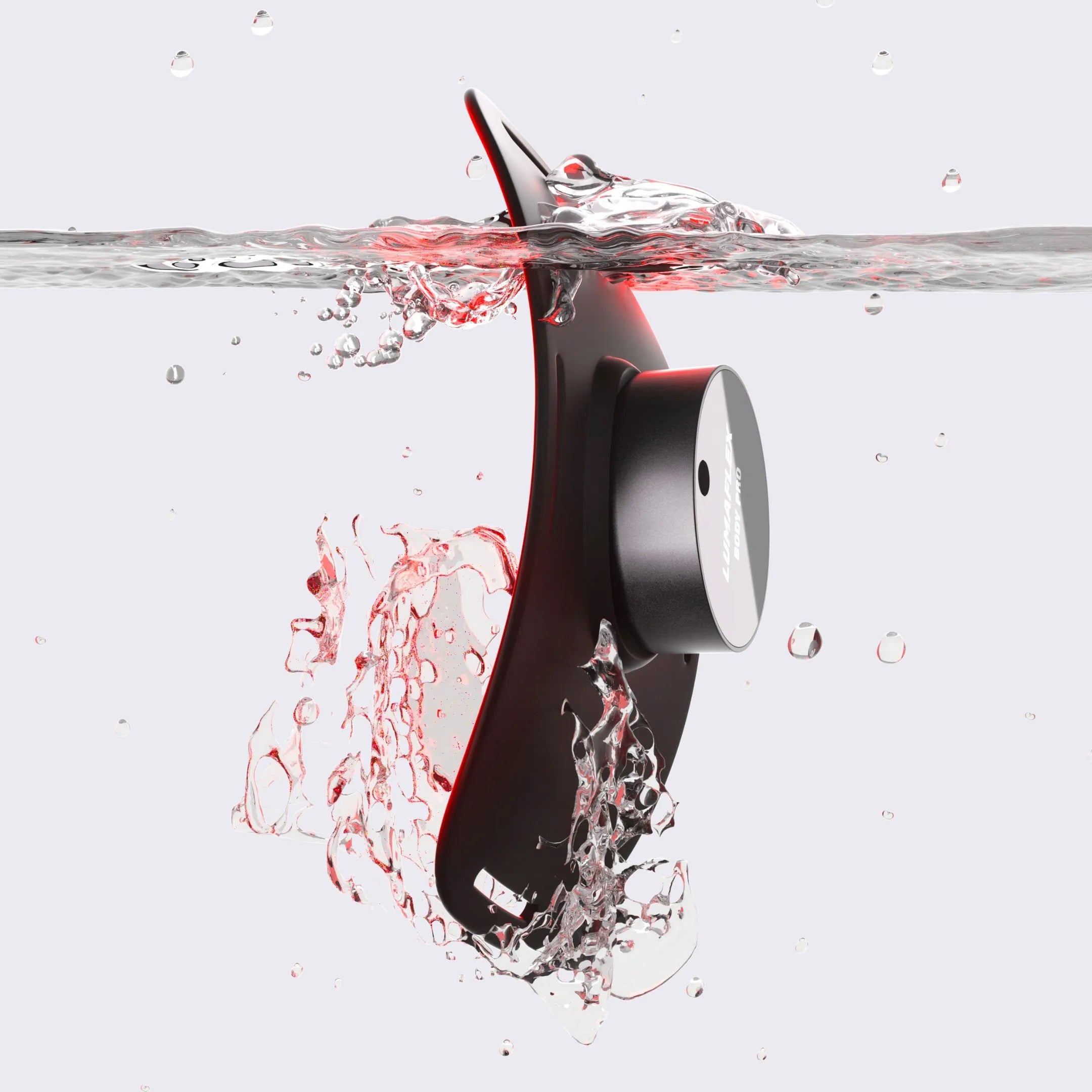
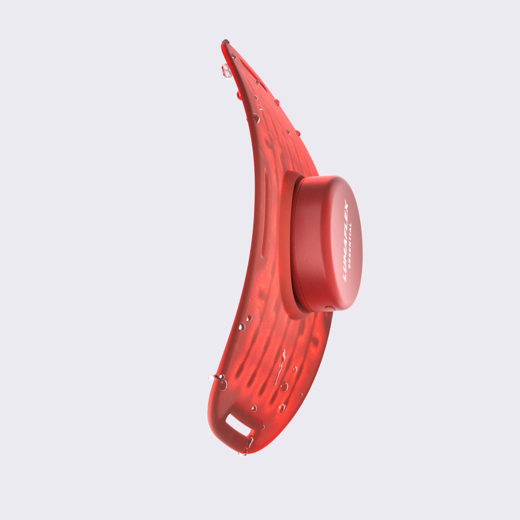
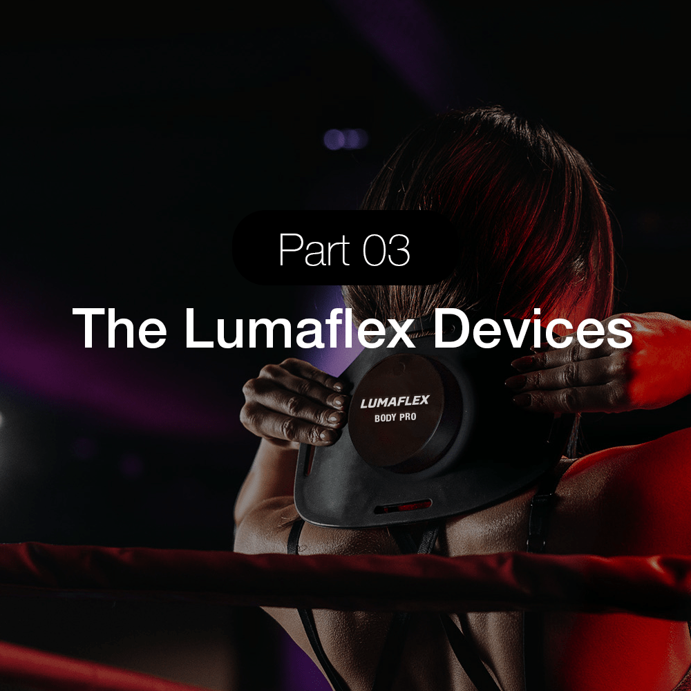
Lumaflex devices use LED-based wearable panels that strap onto areas such as the knee, arm, or shoulder. Their specifications include:
Body Pro: 45 mW/cm² red light irradiance and 30 mW/cm² near-infrared irradiance.
Essential Pro: 35 mW/cm² red light irradiance and 33 mW/cm² near-infrared irradiance, with six wavelengths: 630, 660, 810, 850, 904, and 1064 nm.
Essential: Red light paired with 850 nm near-infrared light.
Several wavelengths offered by Lumaflex devices fall within the near-infrared range investigated in human tendinopathy trials. However, those trials used laser diodes applied to specific treatment points, while Lumaflex devices use LEDs in wearable panels. The wavelength is only one part of a treatment protocol. Irradiance, dose, treatment area, exposure time, and light delivery method also influence how the treatment is delivered.
Lumaflex devices are FDA 510(k) cleared for pain management. The tendon studies discussed in this article provide research context for photobiomodulation and tendon recovery, but they do not establish a separate FDA-cleared indication for tendon repair.

The Potential of Red Light Therapy for Tendon Repair
Research into photobiomodulation (PBM) has examined how red and near-infrared light may influence several processes involved in tendon recovery. Animal studies have reported changes in inflammatory markers, collagen organization, and the amount of force tendons can withstand before failure. Human clinical trials have also reported improvements in pain, physical function, and strength, particularly when PBM is combined with exercise-based rehabilitation for chronic tendinopathy. Clinical research following surgical tendon repair is extending this work to structural tendon injuries.
These findings place PBM within a broader approach to tendon rehabilitation, where progressive loading remains central to recovery. Light-based treatment is being investigated as a complementary approach that may influence cellular activity and tissue remodeling alongside established rehabilitation methods. Understanding the treatment parameters is equally important. Wavelength, irradiance, dose, exposure time, and treatment schedule all shape how light is delivered and help explain the differences between research protocols and devices.
As research continues, these factors provide a practical framework for understanding the potential role of red light therapy in tendon recovery and evaluating devices designed for photobiomodulation.