904nm vs 1064nm: Which Light Reaches the Brain?

Why Most Red Light Therapy Doesn't Reach the Brain
If you have looked into photobiomodulation (PBM) to promote brain health, chances are high that you have seen that the majority of available devices fall in the 810 nm to 850 nm range. This range has been well explored and proven highly effective for various uses. However, for the case of brain tissue and especially for the cortex, the difference is noticeable.
Red light therapy using 904 nm and 1064 nm wavelengths in brain applications is a more focused method of transcranial photobiomodulation. The interaction of NIR with tissue is different from other wavelengths, and studies on this subject, especially on 1064 nm, have increased both in number and scope.
This article examines both wavelengths in detail: their penetration physics, their distinct mechanisms, what the current evidence actually shows, and how they compare to the standard NIR range most people are familiar with.
- 1. Why Most Red Light Therapy Doesn't Reach the Brain
- 2. Light Penetration 101: Why Wavelength Matters
- 3. What Makes 904nm Different?
- 4. 1064nm: The Deepest Studied NIR Wavelength for the Brain
- 5. How Light Reaches and Affects Brain Tissue
- 6. 904nm vs 1064nm: Key Differences
- 7. What the Research Actually Shows
- 8. Who Might Benefit Most?
- 9. Multi-Wavelength Devices and How 904nm + 1064nm Are Used
- 10. Bringing It Together
- 11. Frequently asked questions (FAQs)
- 12. References
Light Penetration 101: Why Wavelength Matters
Light interacts with biological tissue in different ways. When the light enters the body, there are three main forces that act on it: absorption, scattering, and transmission. Absorption occurs as a result of interactions with substances in the tissues, such as hemoglobin, melanin, and water. The relationship between these three forces depends greatly on the wavelength.
Researchers refer to the range between roughly 650nm and 1100nm as the optical window. Within this band, light faces minimal obstruction from the dominant tissue absorbers, which allows it to travel further before losing energy. This is why NIR wavelengths are used in therapeutic contexts at all.

Wavelength selection matters significantly when the target is as deep and shielded as the brain. The skull alone presents a multi-layer barrier: skin, subcutaneous fat, bone, and meningeal layers, each with different optical properties. Standard 810nm to 850nm light can cross this barrier to some degree, but energy attenuation is substantial by the time photons approach cortical tissue.
| Key Concept: |
| Think of wavelength as a key. Not because every lock opens, but because tissue layers respond to different frequencies with different levels of resistance. Longer wavelengths within the optical window tend to face less absorption, which translates to deeper reach. |

What Makes 904nm Different?
904 nm falls just above the usual range of NIR used for therapy, and this affects the way that light behaves in tissue. With this wavelength, light absorption by water is still low, while light absorption by hemoglobin has decreased dramatically in comparison with shorter NIR wavelengths. This allows more free movement of light within tissue..
One distinguishing feature of 904nm devices is that they are often delivered as pulsed laser light rather than continuous wave. Pulsed delivery allows higher peak power at lower average intensity, which can reduce thermal buildup while maintaining photon flux in target tissue. This matters for safety and for reaching deeper structures without causing surface-level discomfort.
904nm has a reasonably established record in pain management and musculoskeletal applications. Its use in neural modulation contexts is less documented at the brain level specifically, but its peripheral nerve effects and depth advantage over 810nm make it a meaningful part of a multi-wavelength therapeutic approach.
| Important Nuance: |
| 904nm has a smaller body of brain-specific research compared to 1064nm. Claims about direct cortical effects should be interpreted cautiously. Its value in brain-focused protocols may lie more in systemic support and synergy with other wavelengths than in direct transcranial action. |
1064nm: The Deepest Studied NIR Wavelength for the Brain
1064nm is where the transcranial photobiomodulation research becomes most compelling. There have been several studies conducted by peers, based on which it has been proved that light of wavelength 1064nm can reach a depth of about 5mm of the cortex under natural tissue conditions. This depth assumes significance when it is considered that the prefrontal cortex lies within this depth.
One important factor contributing to the penetration ability of the wavelength lies in its location in the electromagnetic spectrum. In other words, 1064nm occurs in a range where water absorption starts to rise but is not so high that substantial amounts of light are unable to penetrate deeply into tissues. On the other hand, the scattering coefficient falls with increasing wavelengths.
Published research has linked 1064nm transcranial application with improvements in working memory tasks, reaction time, and measures of cognitive efficiency in healthy adults. These are human studies with measurable outcomes, not just theoretical models, which is why 1064nm has become the benchmark wavelength for serious transcranial PBM investigation.
How Light Reaches and Affects Brain Tissue
Understanding the mechanism behind photobiomodulation is essential for evaluating any wavelength-specific claim. The process begins at the mitochondrial level, with a protein called cytochrome c oxidase (CCO).
The CCO Pathway: From Photon to Cellular Response
CCO is the end product of the mitochondrial electron transport chain. It responds to light over a wide range of near-infrared wavelengths, and once activated, triggers a series of cascading responses thought to be responsible for most of the benefits seen from PBM.

Here is the general sequence, as current research describes it:
1. Photons are absorbed by CCO in mitochondria within neural cells
2. Increased CCO activity drives higher ATP (adenosine triphosphate) production
3. Nitric oxide, which had been inhibiting CCO, is released
4. Released nitric oxide dilates blood vessels, improving local cerebral circulation
5. Reduced oxidative stress and inflammation follow as mitochondrial function normalizes
The net effect is a more metabolically active, better-oxygenated neural environment. For a brain recovering from fatigue, metabolic stress, or suboptimal function, that shift can be meaningful.
This mechanism is not wavelength-exclusive, but the efficiency of CCO activation varies with wavelength. 1064nm in particular aligns with one of CCO's absorption peaks, which may partially explain its stronger research outcomes at the transcranial level.
904nm vs 1064nm: Key Differences
Both wavelengths operate within the optical window and share the core PBM mechanism through CCO. But their practical profiles are distinct enough to matter when choosing or evaluating a device.
| Feature | 904nm | 1064nm |
| Penetration depth | Deep peripheral & nerve tissue | Deepest cortical (up to ~5mm) |
| Delivery method | Pulsed laser | Continuous or pulsed |
| Research base | Moderate | Strongest for transcranial PBM |
| Primary role | Synergistic support | Primary brain targeting |
| Water absorption | Low | Slightly higher, still viable |
The distinction is not simply that one is 'better' than the other. 904nm has an advantage in terms of depth when compared to shorter NIR in the peripheral and nerve tissues, and there is adequate evidence suggesting its influence on a systemic level. On the other hand, 1064nm has strong evidence for transcranial transmission and cortical interaction.
In a multi-wavelength protocol, both could be used for complementary purposes: 904nm to assist in nerve and vascular regulation in the surrounding tissue, whereas 1064nm would generate the deeper cortex stimulation.
What the Research Actually Shows
It is important to be clear-eyed about where the science currently stands. The evidence base for transcranial photobiomodulation is genuinely encouraging, but it is also early-stage in many respects.
Human Studies
A number of controlled studies have employed the use of 1064nm radiation over the forehead and pre-frontal areas. The subjects displayed improvement in terms of working memory functioning, sustained attention, and faster processing times than the control group, which was subjected to placebo.
Animal Studies
Animal models have provided more mechanistic detail, including direct measurement of ATP increases and nitric oxide changes in neural tissue following NIR exposure. Monte Carlo simulations have been used to model light propagation through realistic skull geometry, providing depth estimates that align with what human studies suggest clinically.

Honest Assessment of the Evidence
In the scientific literature relating to brain performance, about 60% to 70% of published articles have reported positive results. Most of the experiments used small numbers of subjects and diverse procedures without standardization. In addition, there is a dearth of large-scale, randomized controlled studies that use uniform parameters and measurements. Early results show promise, but more research is needed before reaching the standard of well-proven medicine.
| Research Reality |
| Photobiomodulation for brain health is a promising, actively developing field. The evidence is substantive enough to take seriously, but it should be understood as emerging science rather than settled clinical fact. |
Who Might Benefit Most?
The population showing the most interest in transcranial PBM falls into several broad categories, each with different reasons for exploring this approach.
Cognitively Active Adults
People working in high-demand cognitive roles or those focused on maintaining mental performance as they age represent a significant portion of PBM users. Early research suggests benefits in working memory and processing speed, which are naturally of interest to this group.
Neurological Recovery
Individuals in post-injury or post-illness recovery, particularly those dealing with traumatic brain injury or stroke-related cognitive effects, appear in several of the more focused clinical studies. Research suggests that mitochondrial support and improved cerebral blood flow may be particularly relevant in recovery contexts, though this remains an area requiring much more rigorous investigation.
Biohackers and Wellness Practitioners
Those already familiar with the broader landscape of photobiomodulation often approach deeper-wavelength devices as a logical progression from surface-level red light therapy. This audience tends to be well-informed about the limitations of current evidence and is comfortable working with emerging protocols.
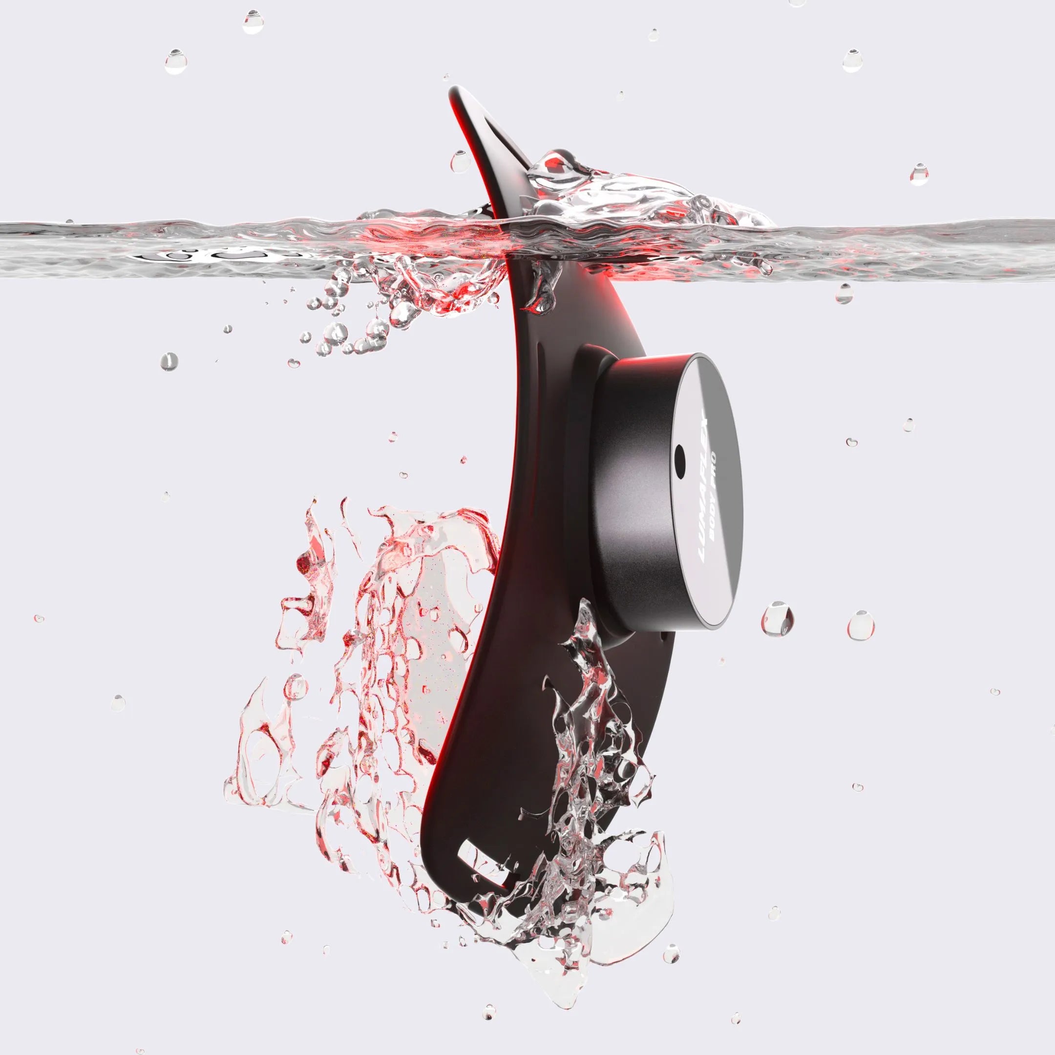
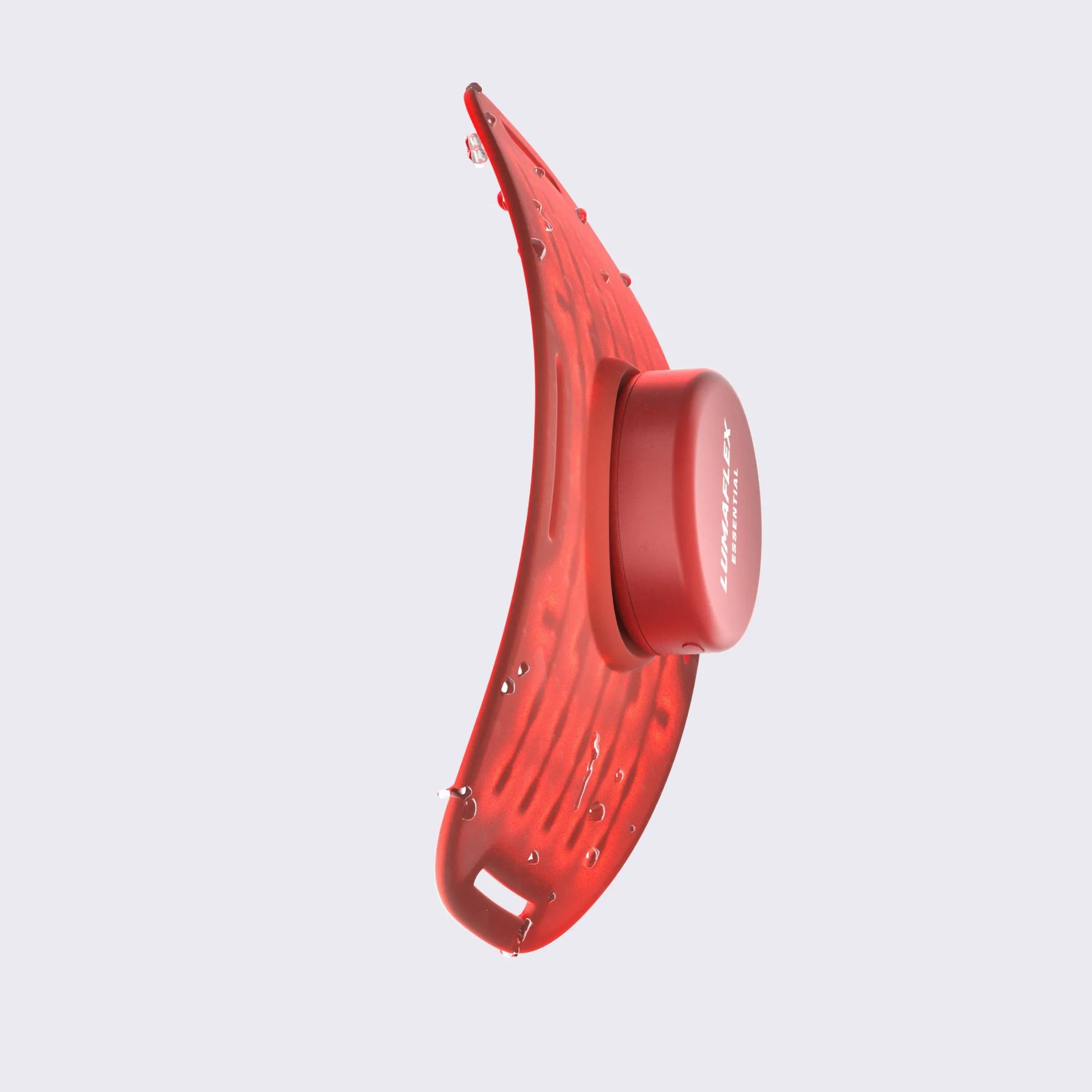
Multi-Wavelength Devices and How 904nm + 1064nm Are Used
Effective delivery of transcranial PBM therapy goes beyond choosing the proper wavelength. The power density, duration of exposure, pulse frequency, and positioning will all affect light penetration as well as biological response.
Several multi-wavelength devices, such as Lumaflex Essential Pro, also use both 904nm and 1064nm frequencies in their overall spectrum range. It is because using both frequencies at the same time ensures that a more holistic treatment strategy can be achieved. This means that while the frequency of 904nm will focus on nerve and vascular tissues, that of 1064nm will concentrate on cortical tissues.
For anyone evaluating a device for brain-focused applications specifically, the presence of 1064nm is the most evidence-supported specification to look for. The delivery parameters, including whether pulsing is available and what the output power is, matter considerably as well.
Bringing It Together
904 nm and 1064 nm are important in brain research because they have the ability to access areas that NIR wavelengths at lower nanometers cannot reach, and they have the ability to trigger a known biological process that has tangible impacts on brain metabolism and circulation.
1064 nm has the greatest research backing regarding transcranial PBM therapies because of its high penetration depth, compatibility with the absorption peak of CCO, and cognitive results in human tests. The 904 nm wavelength also plays a useful auxiliary role, especially with regards to peripheral nerve and vascular tissue, where it might prove beneficial in a multi-wavelength approach.
The truth is that the field is progressing fast, initial signs are promising, and there is a solid foundation in the biology of mitochondria. The thing that is missing is the scope of randomized trials necessary for clinical confidence. This does not detract from what we know so far, but it is something to keep in mind.
If you have already worked with photobiomodulation technology, the issue of choosing between devices that only work at a superficial level through near-infrared light and the kind that can truly affect brain tissues will be your first step.
What wavelength reaches the brain?
1064nm has the strongest evidence for cortical penetration, reaching approximately 5mm into brain tissue under optimal conditions.
Does red light penetrate the skull?
Yes, partially. Near-infrared light can pass through bone and scalp, though a significant portion is absorbed or scattered before reaching deeper structures.
Is 904nm effective for brain therapy?
904nm shows indirect support through neural and peripheral tissue effects. Brain-specific research is still emerging and limited compared to 1064nm.
How deep does NIR light penetrate?
Penetration varies by tissue density and wavelength. Estimates range from 5mm to 10mm for NIR light in cortical tissue, depending on the study model used
Is photobiomodulation clinically proven?
Results are early-stage but encouraging. Multiple human studies show measurable cognitive improvements, though large-scale randomized trials are still needed.
References
Barrett, D. W., & Gonzalez-Lima, F. (2013). Transcranial infrared laser stimulation produces beneficial cognitive and emotional effects in humans. Neuroscience, 230, 13–23. https://doi.org/10.1016/j.neuroscience.2012.11.016
Cassano, P., Petrie, S. R., Hamblin, M. R., Henderson, T. A., & Iosifescu, D. V. (2016). Review of transcranial photobiomodulation for major depressive disorder: Targeting brain metabolism, inflammation, oxidative stress, and neurogenesis. Neurophotonics, 3(3), 031404. https://doi.org/10.1117/1.NPh.3.3.031404
Gonzalez-Lima, F., Barksdale, B. R., & Rojas, J. C. (2014). Mitochondrial respiration as a target for neuroprotection and cognitive enhancement. Biochemical Pharmacology, 88(4), 584–593. https://doi.org/10.1016/j.bcp.2013.11.010
Hamblin, M. R. (2016). Shining light on the head: Photobiomodulation for brain disorders. BBA Clinical, 6, 113–124. https://doi.org/10.1016/j.bbacli.2016.09.002
Hamblin, M. R. (2017). Mechanisms and mitochondrial redox signaling in photobiomodulation. Photochemistry and Photobiology, 94(2), 199–212. https://doi.org/10.1111/php.12864
Hennessy, M., & Hamblin, M. R. (2017). Photobiomodulation and the brain: A new paradigm following in the footsteps of low-level light therapy. Journal of Optics, 19(1), 013003. https://doi.org/10.1088/2040-8986/19/1/013003
Jagdeo, J. R., Adams, L. E., Brody, N. I., & Siegel, D. M. (2012). Transcranial red and near infrared light transmission in a cadaveric model. PLOS ONE, 7(10), e47460. https://doi.org/10.1371/journal.pone.0047460
Lapchak, P. A., & De Taboada, L. (2010). Transcranial near infrared laser treatment (NILT) increases cortical adenosine-5′-triphosphate (ATP) content following embolic strokes in rabbits. Brain Research, 1306, 100–105. https://doi.org/10.1016/j.brainres.2009.10.022
Naeser, M. A., Zafonte, R., Krengel, M. H., Martin, P. I., Frazier, J., Hamblin, M. R., Knight, J. A., Meehan, W. P., & Baker, E. H. (2014). Significant improvements in cognitive performance post-transcranial, red/near-infrared light-emitting diode treatments in chronic, mild traumatic brain injury: Open-protocol study. Journal of Neurotrauma, 31(11), 1008–1017. https://doi.org/10.1089/neu.2013.3244
Nawashiro, H., Wada, K., Nakai, K., & Sato, S. (2012). Focal increase in cerebral blood flow after treatment with near-infrared light to the forehead in a patient in a persistent vegetative state. Photomedicine and Laser Surgery, 30(4), 231–233. https://doi.org/10.1089/pho.2011.3044
Rojas, J. C., & Gonzalez-Lima, F. (2013). Neurological and psychological applications of transcranial lasers and LEDs. Biochemical Pharmacology, 86(4), 447–457. https://doi.org/10.1016/j.bcp.2013.06.012
Salehpour, F., Mahmoudi, J., Kamari, F., Sadigh-Eteghad, S., Rasta, S. H., & Hamblin, M. R. (2018). Brain photobiomodulation therapy: A narrative review. Molecular Neurobiology, 55(8), 6601–6636. https://doi.org/10.1007/s12035-017-0852-4
Tedford, C. E., DeLapp, S., Jacques, S., & Anders, J. (2015). Quantitative analysis of transcranial and intraparenchymal light penetration in human cadaver brain tissue. Lasers in Surgery and Medicine, 47(4), 312–322. https://doi.org/10.1002/lsm.22343
Tian, F., Hase, S. N., Gonzalez-Lima, F., & Liu, H. (2016). Transcranial laser stimulation improves human cerebral oxygenation. Lasers in Surgery and Medicine, 48(4), 343–349. https://doi.org/10.1002/lsm.22471
Wang, X., Tian, F., Soni, S. S., Gonzalez-Lima, F., & Liu, H. (2016). Interplay between up-regulation of cytochrome-c-oxidase and hemoglobin oxygenation induced by near-infrared laser. Scientific Reports, 6, 30540. https://doi.org/10.1038/srep30540