Red Light Therapy for Brain Health: Complete Hz & Wavelength Guide (10Hz–40Hz)

Red light therapy for brain health.
Brain health, what has previously been a niche field of study, is now one of the most widespread research interests in the current day; with people increasingly looking to sustain cognitive health and prevent further progression of neurological disorders. Research into neurological injury, from conditions like stress, lack of sleep and physical trauma is developing at an incredible pace and while research has mostly focused on diet and exercise there is now an increasing amount of literature which suggests that light may be integral.
Photobiomodulation (PBM) is the clinical term for using specific wavelengths of red and near-infrared light to produce biological effects in tissue. When applied to the scalp, it becomes transcranial photobiomodulation, a non-invasive technique that delivers light energy to brain tissue through the skull. What has drawn researchers' attention is the range of effects this appears to produce: improved mitochondrial function in neurons, increased blood flow to cortical regions, and measurable changes in cognitive performance across a variety of populations.
Almost every one learning about red light therapy on the brain will learn about the wavelengths. That near-infrared light at 810nm can travel through the skull to the cortical tissues. This is true, and important. What is less discussed is the other variable impacting results; frequency, or Hz. The speed with which your device pulses its light is not an inconsequential specification; it is a biological signal that is communicated to the rhythms of the brain and what the therapy actually does is determined by what Hz you use to achieve your objective.
This guide covers the mechanisms, the research, the wavelengths, the specific frequency values, and how to put them together into a practical protocol.
This article is for educational purposes and reflects publicly available research on photobiomodulation. It is not medical advice. Always consult a qualified healthcare professional for any medical condition.
- 1. How Red Light Therapy Affects the Brain
- 2. What the Research Shows
- 3. Wavelengths for Brain Health
- 4. Red Light Therapy Frequencies for Brain Function
- 5. Building a Brain Health Protocol
- 6. Benefits of Red Light Therapy for Brain Health
- 7. Safety and Practical Considerations
- 8. How Lumaflex Supports Brain Health Protocols
- 9. Final Thoughts: Frequency and Wavelength Together
- 10. Related Reading
- 11. References
How Red Light Therapy Affects the Brain
The effects of transcranial PBM are not produced by a single mechanism. Several interconnected biological processes respond to photonic stimulation, and understanding them helps explain why different frequency and wavelength combinations produce different outcomes.

Mitochondrial Function and ATP Production
The chief mechanism behind PBM therapy on any body tissues is the mitochondria, which are cells in charge of ATP generation. This ATP is the chief energy source of any biological cell. Cytochrome c oxidase is an enzyme located within the respiratory chain of the mitochondria that absorbs photons in the red and near-infrared spectrum. When such absorption happens, it enhances ATP generation and decreases oxidative stress.
The neurons are one of the most energy-intensive types of cell. Mitochondrial energy production can be impaired when brain tissue is exposed to different stress factors such as trauma, infection, aging, and metabolic disorders. Transcranial photobiomodulation increases the efficiency of mitochondrial processes in neurons within the cortex and enhances the rate of oxygen uptake and ATP generation in dysfunctional tissue.
Cerebral Blood Flow
Perhaps one of the more consistently noted effects of transcranial PBM is an increase in local blood flow within the brain. This is mediated through nitric oxide signaling. After absorbing photons, cells will produce and release nitric oxide, causing local vasodilation in order to improve blood flow to the area. Delivery of blood carries with it oxygen and glucose to the brain, which will assist with cognitive function as well as recovery.
Several imaging studies have confirmed this effect using functional near-infrared spectroscopy and other blood flow measurement tools. The increases in oxygenated hemoglobin in frontal and prefrontal regions following transcranial PBM sessions have been documented in both healthy adults and in populations with cognitive impairment.
Neuroinflammation Reduction
Neuroinflammation plays a role in many brain-related issues, such as post-concussion syndrome and neurodegenerative disorders. Microglia, which act as the brain's own immune cells, are capable of becoming constantly activated, causing damage to the neurons nearby and emitting inflammation signals. It has been proven that photobiomodulation influences microglia and decreases the inflammatory effect on the brain's tissue.
This anti-inflammatory effect is part of why transcranial PBM has attracted attention in the context of traumatic brain injury, where neuroinflammation is a major driver of ongoing symptoms long after the initial injury.
Neural Signaling and Synaptic Support
Apart from its effect in energy production and inflammation, there is evidence that PBM impacts neurogenesis and synaptogenesis. In animals, it has been noted that there is an increase in the development of hippocampal cells following exposure to PBM, and there are improved learning and memory tasks. Although it may not be prudent to transfer animal results directly to humans, this can provide a biological rationale for the effects seen in cognitive function, especially memory and executive function, in humans.
What the Research Shows
While the body of literature on transcranial photobiomodulation is relatively small compared to those on drugs, there have been several rigorously designed studies which yielded findings that cannot be easily overlooked.
TBI Recovery and Cognitive Improvement: Naeser et al. (2014, 2016)
The studies that have gained wide acceptance in this regard include those carried out by Dr. Margaret Naeser and associates from Boston University School of Medicine and the VA Boston Healthcare System. In one open-protocol study conducted in 2014, which was published in the Journal of Neurotrauma, 11 patients suffering from mTBI were subjected to 18 sessions of transcranial LED therapy within a period of six weeks. All the individuals who underwent the procedure suffered from long-term neuropsychological impairment due to mTBI, for a duration ranging from 10 months to as much as 8 years following the incident. Significant improvement was noted on neuro-psychological assessments of executive function, verbal memory, and sleep, as well as social and professional performance.
A review published in Photomedicine and Laser Surgery in 2016 by Naeser et al. further elucidated on this topic and described the mechanisms involved in this process: there is increased ATP production by mitochondria, which causes nitric oxide to be released for improved cerebral blood flow and reduces neuroinflammation within the tissue. The paper also highlighted how 10 Hz pulsation had superior results compared to continuous waves in an animal model in previous experiments.
10 Hz Pulsed Delivery and Neuroprotection
One review published in Photomedicine and Laser Surgery in 2015 assessed the scientific knowledge on the use of transcranial PBM treatment for TBI patients by compiling the research data collected both from experiments conducted on animals and initial human trials. Among the interesting results that emerged from research on animal subjects was one which stated that a pulsed laser with a frequency of 10 Hz was better than either continuous wave lasers or lasers which pulsed at 100 Hz in treating injured brains. The idea here is based on resonance; because the pulse frequency used in the treatment coincides with that produced by the hippocampus, a part of the brain associated with memory, it stimulates or strengthens the latter's electrical waves.
Alzheimer's Disease and Cognitive Decline
In an article that appeared in the Journal of Alzheimer's Disease, the effects of transcranial PBM were evaluated for the treatment of probable CTE, which is a progressive disease of the brain caused by repeated head injuries. Four former NFL players underwent 18 PBM treatments, with 810 nm LED pulses delivered at a frequency of 40 Hz. Positive results were found regarding cognitive function, behavior, and mood, as well as brain imaging that indicated improved neural communication and metabolism.
This is one of the most direct pieces of evidence for the specific utility of the 40 Hz frequency in brain health applications. The 40 Hz gamma frequency was chosen intentionally, based on prior research linking gamma oscillations to cognitive processing and the observation that gamma power is reduced in neurodegenerative conditions.
Broader Cognitive Benefits Across Populations
An article titled “Cognitive Impairments After Transcranial PBM Therapy” which appeared in Ageing Research Reviews in 2022 systematically reviewed 35 clinical trials on transcranial PBM therapy and concluded that all 35 clinical trials demonstrated positive results with respect to cognitive functioning. In particular, 100% of clinical trials involving people with memory problems, cognitive impairments or dementia demonstrated positive results. For clinical trials involving TBI patients, 87.5% showed positive results.
These are not insignificant results. An intervention with such consistency of benefits across different populations and measures of cognition, one that is also non-invasive and safe, warrants careful consideration by any researcher interested in brain function.
Wavelengths for Brain Health
Penetration through the skull is dependent on wavelengths with enough penetration capability. It is not always the case that any wavelength of red and near infrared light would suffice, as different wavelengths would result in access to different tissue layers.
810 nm and 850 nm: The Primary Brain Wavelengths
The 810nm wavelength is located in one of the absorption peaks of cytochrome C oxidase and is therefore one of the wavelengths most bioactive in PBM. The majority of the human trials conducted with transcranial PBM have utilized the 810nm wavelength as the key wavelength, which also features prominently in the literature with regard to cognitive/neurological use. While having the same characteristics as far as tissue penetration is concerned, the 850nm wavelength is frequently employed with the 810nm wavelength in various studies and clinical cases.
904 nm and 1064 nm: Deeper Penetration for More Intensive Protocols
In cases where the therapy focuses on more sub-cortical brain structures, or when the individual is interested in having all the photons delivered to the cortical layers, 904 nm and 1064 nm provide extra penetration power. According to studies done at MIT’s McGovern Institute, the delivery of 1064 nm light waves across the skull to the pre-frontal cortex resulted in enhanced visual working memory capacity among healthy adults.
In a full-spectrum device like the Lumaflex Essential Pro, which includes 630nm, 660nm, 810nm, 850nm, 904nm, and 1064nm, users can deliver a complete wavelength range covering everything from surface tissue to deeper cortical and sub-cortical regions in a single session. For brain applications, the 810-1064nm range is the working stack.
Why Penetration Depth Matters
The skull reduces photon delivery significantly. Estimates from imaging studies suggest that roughly 1-3% of surface-applied near-infrared light reaches cortical tissue. This is why power density, session duration, and wavelength selection all matter more for brain applications than for surface tissue like skin or muscle. Using wavelengths with strong skull penetration, combined with consistent repeated sessions, is what allows photon energy to accumulate at therapeutically relevant levels in cortical tissue.
Red Light Therapy Frequencies for Brain Function
This is where most red light therapy content stops being useful. Devices either offer no frequency control, or they list preset modes without explaining what the frequency settings are or why they matter. For brain applications specifically, frequency is not a secondary consideration. It is central to how the therapy interacts with the brain's own electrical rhythms.
| Hz Setting | Range Band | Brain Effects | Best Protocol Use |
| 1-5 Hz | Reset & Restore | Parasympathetic activation, sleep prep, deep relaxation | Wind-down, evening recovery, stress reduction |
| 7-8 Hz | Calm & Focus | Alpha-adjacent rhythms, nervous system calming, gentle mood support | Post-workout brain recovery, anxiety reduction |
| 10 Hz | Calm & Focus | Focus support, mood regulation, surface neural stimulation; most studied for PBM | Daily cognitive maintenance, skin + scalp, open TBI research protocols |
| 40 Hz | Activate & Energize | Gamma wave entrainment, cognitive activation, memory, neural synchrony | Cognitive performance sessions, brain health stack, CTE-adjacent research |
| 80-100 Hz | Activate & Energize | Deep neural stimulation, chronic recovery, nociceptive modulation | Post-injury support, deep recovery, intensive protocol days |
| Continuous | Baseline / CW | Baseline photon delivery without timing signal, broad cellular support | Introductory sessions, general wellness, stacking with pulsed runs |
The Concept of Neural Entrainment
The brain is an organ of electricity. Neurons fire together in synchronicity at particular frequencies. These firing patterns show up as brainwaves on an EEG and relate to various states of mind. Delta waves are seen between 0.5 Hz and 4 Hz while someone is sleeping deeply. Theta waves are seen between 4 Hz and 8 Hz while someone is in light sleep and relaxed concentration. Alpha waves are seen between 8 Hz and 12 Hz while someone is awake and relaxed.
Neural entrainment refers to the synchronization between neural oscillations and any periodic external stimulus. If the light pulses occur at a rate equal to one of the biological brain wave bands, then the brain will entrain itself with this rhythm. This effect is not hypothetical but has been empirically studied in many PBM experiments using the EEG test. The gamma brain waves with the frequency of 40 Hz are best known in such entrainment.
Why 10 Hz Stands Out in the Research
10 Hz is the most studied frequency in transcranial PBM research, appearing in multiple clinical protocols and animal studies. Its position within the alpha brainwave range, and its resonance with hippocampal electrical activity, gives it particular relevance for memory, focus, and mood regulation applications. The 2015 review examining PBM for TBI found that 10 Hz pulsed delivery outperformed both continuous wave and higher-frequency pulsing in animal injury models, with hippocampal resonance cited as the likely mechanism.
For everyday use, 10 Hz sessions are typically used for daytime cognitive support, focus before demanding cognitive tasks, mood regulation, and as the base frequency in a brain health maintenance protocol.
Why 40 Hz Is the Most Consequential Brain Frequency
The 40 Hz frequency is considered to be within the brain wave frequency category known as the gamma brain wave range. Of all the individual frequencies studied in terms of brain functions, there is no doubt that this is the most widely studied. The gamma brain waves are responsible for cognitive functions like working memory and attention, among others. The key factor here is that the levels of gamma brain waves are significantly lowered in patients with diseases like Alzheimer’s, Parkinson’s, and those who have suffered brain injury.
Findings from researchers at MIT and elsewhere have established that the use of flicker stimulation to drive 40 Hz oscillations results in altered brain function and in some cases decreases amyloid plaque and tau protein deposition in animals. The findings reported in the 2023 study by Naeser et al. involving retired NFL players who received LED lights at 810nm stimulated at 40 Hz frequency showed improved cognition with brain connectivity changes.
In practice: 40 Hz is the frequency that should be used when cognitive activation is the target. This includes anything before work, study sessions, or any period of time where higher brain function is desired.
The Role of Continuous Mode
There is also the continuous wave delivery approach, whereby the light is delivered continuously without pulsation. Many of the first experiments performed using transcranial PBM were conducted with the continuous wave approach and they gave some positive outcomes. The continuous wave mode provides constant photon energy without an accompanying time signal, and thus could be helpful in the beginning phases, as well as in other scenarios where a general boost in brain wellness is needed, including as a base of a stacked protocol with pulsations.

Building a Brain Health Protocol
The research points toward a few clear principles for structuring a brain-focused photobiomodulation protocol. Wavelength determines where the light goes. Frequency determines what it tells the tissue to do. Consistency determines whether the effects accumulate. Below are protocol structures based on the research and frequency logic covered in this article.
| Protocol | Hz | Wavelength | Goal |
| Foundational Brain Stack | 10 Hz + 40 Hz alternating | 810 nm / 850 nm | Daily cognitive support, focus, neural health baseline |
| Cognitive Performance | 40 Hz | 810 nm | Gamma entrainment, pre-work or study session activation |
| Recovery & Wind-Down | 7-8 Hz then 1-5 Hz | 850 nm | Post-cognitive load, evening session, nervous system reset |
| Advanced Deep Recovery | 80-100 Hz | 904 nm / 1064 nm | Post-injury support, intensive recovery days, chronic conditions |
| Broad Wellness Baseline | Continuous mode | 810 nm / 850 nm | New users, maintenance protocol, stacking foundation |

Foundational Brain Stack
For most individuals looking to introduce a brain wellness protocol into their routine, the foundational stack should be used to start with: 10 Hz and 40 Hz alternatively, utilizing wavelengths of either 810 nm or 850 nm. The reason being that this protocol will cover both memory and mood stimulation from the 10 Hz wave and the brain activation from the 40 Hz wave.
Session duration of 10 minutes per placement is the most common protocol in the research literature. Consistency over weeks, rather than longer individual sessions, produces the cumulative effects observed in clinical studies.
Advanced Stack with Deeper Wavelengths
Users with deeper recovery goals, or those dealing with chronic neurological challenges, can layer in 904nm and 1064nm wavelengths alongside higher Hz settings in the 80-100 range. The deeper wavelengths increase photon delivery to sub-cortical tissue and bone-adjacent structures. This is a more intensive protocol and should be approached progressively rather than started at full intensity.
Benefits of Red Light Therapy for Brain Health
Based on the mechanisms and research described above, here is a summary of the specific outcomes that transcranial PBM has been associated with in clinical literature.
Focus and Mental Clarity
Multiple studies have reported improved attention, concentration, and processing speed following transcranial PBM. The increase in cerebral blood flow and ATP production in prefrontal cortical regions provides a plausible biological explanation. For healthy adults, this tends to manifest as sharper focus during cognitively demanding tasks and reduced mental fatigue.
Memory and Cognitive Performance
The most consistent finding across the research is improved memory, particularly working memory and verbal memory. The hippocampal resonance at 10 Hz, and the gamma-related improvements at 40 Hz, both point toward mechanisms that support memory encoding and retrieval. The systematic review finding that 100% of studies on populations with cognitive impairment showed positive memory outcomes is particularly notable.
Recovery and Resilience After Injury
It is with regard to TBI, concussion, and post-concussion syndrome that Transcranial PBM has delivered its most striking achievements. Chronic cognitive dysfunction patients participating in the studies carried out by Maeser have displayed improvements following treatment with 18 treatments. The reduction in inflammation in the brain, improvement in mitochondrial efficiency, and increase in cerebral circulation seem to enhance the self-repairing process of the brain in unique ways.
Long-Term Brain Resilience
The neuroprotective angle would seem to be the most promising approach for younger and healthy consumers. Gamma entrainment leading to a decrease in amyloid buildup, the anti-inflammatory properties, and neurogenesis all suggest physiological pathways that may promote brain well-being without merely alleviating symptoms. Although this particular aspect of research is relatively new, the biological feasibility is so convincing that even scientists from prestigious universities such as MIT and Boston University are studying it.
Safety and Practical Considerations
Transcranial photobiomodulation has a strong safety record across the published literature. No serious adverse effects have been reported in any of the human studies reviewed here. The therapy is non-invasive, non-thermal at appropriate power densities, and does not involve ionizing radiation.
Session Duration and Frequency
The most common session duration in research protocols is 10 minutes per placement location. Some studies used up to 20-30 minutes with placement changes, but 10 minutes is the standard starting point and appears to produce measurable effects. For at-home use, once daily or every other day provides sufficient time for cellular responses to accumulate without overexposure.
Consistency Matters More Than Intensity
The pattern in the research is clear: effects accumulate over weeks, not sessions. The cognitive improvements in the Naeser TBI studies emerged after 18 sessions across 6 weeks, not after a single treatment. Approaching brain health PBM as a consistent daily or near-daily practice, rather than an intensive intervention, aligns with how the cellular mechanisms actually work.
Positioning and Application
For transcranial application, the device should be positioned at or near the scalp, over areas corresponding to the intended target. The forehead and prefrontal region is the most commonly used site in research protocols targeting executive function and memory, which maps to the frontal lobe and prefrontal cortex. Some protocols also address the top and sides of the head to cover wider cortical areas.
This article does not constitute medical advice. Red light therapy is not a treatment for any medical condition unless cleared by the relevant regulatory authorities for that specific indication. Consult a qualified healthcare professional before starting any new protocol, particularly if you have a history of neurological conditions.
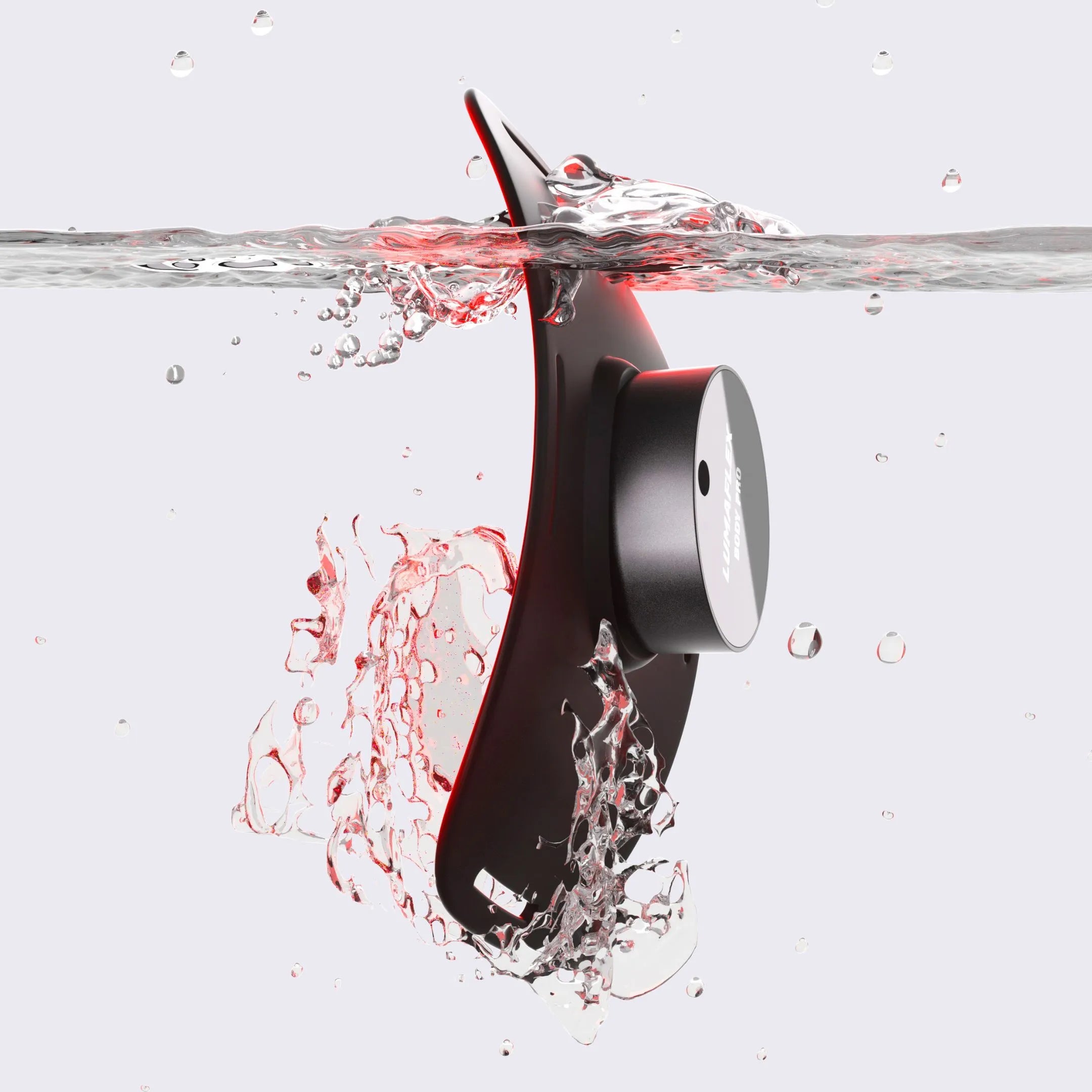
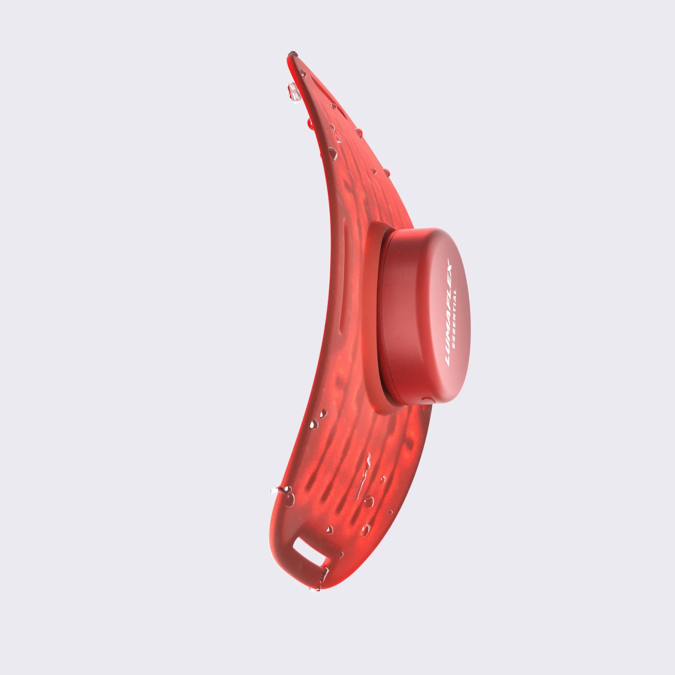
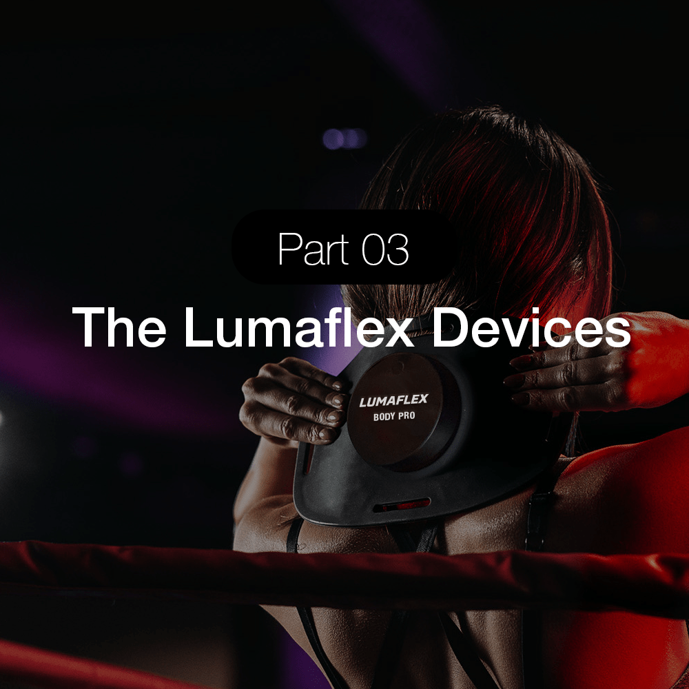
How Lumaflex Supports Brain Health Protocols
One of the most persistent limitations of consumer red light therapy devices is the absence of meaningful frequency control. Most devices offer one or two fixed settings, or broad presets labeled with outcomes like "recovery" or "energy." These categories are too wide to allow the kind of frequency-specific targeting that the research describes.
App-Based Hz Control on the Body Pro and Essential Pro
The Lumaflex Body Pro and Essential Pro both connect to the Lumaflex app, which provides a continuous 1-100 Hz frequency control range. Users can set exactly 10 Hz for a focus session, exactly 40 Hz for a gamma protocol, or exactly 7-8 Hz for a wind-down recovery session, rather than selecting from a small menu of preset modes that may or may not correspond to the frequencies shown to produce specific outcomes in research.
This matters because a device set to a broad "recovery" preset at an unspecified frequency is not the same as a device running at 10 Hz. The research specificity is lost in the translation. Having the ability to select the exact Hz value makes it possible to actually replicate the protocol structures described in published studies.
Six-Wavelength Coverage on the Essential Pro
The six wavelength range offered by The Essential Pro, ranging from 630nm to 1064nm, enables users to experience complete penetration depth capability required for an adequate brain protocol. Wavelengths of 810nm and 850nm offer the delivery of photons into primary cortices. Meanwhile, wavelengths of 904nm and 1064nm offer more profound delivery into brain tissues. Running both simultaneously during a 10-minute session allows the device to provide photon energy at various depths.
The Goal: Precision, Not Just Exposure
The distinction between a precise protocol and general light exposure becomes more important as brain applications are the target, because the mechanisms involved, particularly neural entrainment and frequency-specific mitochondrial response, depend on the timing signal as much as the light itself. The ability to set an exact Hz value is what separates protocol-based therapy from general wellness exposure.
For users who want structured guidance, the Lumaflex Academy included with device purchase covers the science behind frequency settings and how to apply specific protocols, including brain health applications.
Final Thoughts: Frequency and Wavelength Together
Red light therapy for brain health is complicated in that there are many different mechanisms involved, many different wavelengths of light used, and many different frequencies applied, each affecting a distinct part of the process of the light therapy interacting with brain tissue. It is made apparent in the scientific literature that results are dependent upon specificity—the correct wavelength to target light at the correct depth and the correct frequency to deliver the biological message.
10 Hz for maintenance of cognitive function and memory enhancement. 40 Hz for the entrainment of gamma waves and cognitive activity. 810nm and 850nm as primary transcranial wavelengths. 904nm and 1064nm for deep penetration procedures. This is not an arbitrary selection but rather years of scientific study conducted in both healthy individuals, aging individuals, and patients with traumatic brain injuries.
The field is still developing. Larger clinical trials would be useful, but many of the more convincing results come from small sample sizes or even from experiments on animals. However, the reasoning behind these studies seems sensible, their track record of success is good, and the results are sufficiently consistent that the theory should be taken seriously.
For a full breakdown of the frequency ranges used in Lumaflex devices and how to program specific Hz settings in the app, visit the frequency guide.
For the broader context on how Hz and wavelength interact across all applications, see the red light therapy frequency guide at lumaflex.com.
Related Reading
• Red Light Therapy Frequency Guide: What Hz Settings Actually Do to Your Body
• 630nm vs 850nm: Red and Near-Infrared Light Compared
• Discover the Benefits of Red Light at Night for Your Health
• Understanding Light Therapy Color Meanings with Lumaflex
• Red Light Wavelengths Explained by How You’ll Actually Use Them
• Red Light Therapy for Pain 101: Benefits and How It Works
• Transcranial Photobiomodulation: What the Research Actually Shows
References
Ando, T., Xuan, W., Xu, T., Dai, T., Sharma, S. K., Kharkwal, G. B., Huang, Y., Wu, Q., Whalen, M. J., Sato, S., Obara, M., & Hamblin, M. R. (2011). Comparison of Therapeutic Effects between Pulsed and Continuous Wave 810-nm Wavelength Laser Irradiation for Traumatic Brain Injury in Mice. PLoS ONE, 6(10), e26212. https://doi.org/10.1371/journal.pone.0026212
Lee, T., & Chan, A. S. (2023). Photobiomodulation may enhance cognitive efficiency in older adults: a functional near-infrared spectroscopy study. Frontiers in Aging Neuroscience, 15, 1096361. https://doi.org/10.3389/fnagi.2023.1096361
Ma, Y., & Han, Y. (2024). Targeting the brain’s glymphatic pathway: A novel therapeutic approach for cerebral small vessel disease. Neural Regeneration Research, 21(2), 433–442. https://doi.org/10.4103/nrr.nrr-d-24-00821
Naeser, M. A., Martin, P. I., Ho, M. D., Krengel, M. H., Bogdanova, Y., Knight, J. A., Yee, M. K., Zafonte, R., Frazier, J., Hamblin, M. R., & Koo, B. (2016). Transcranial, Red/Near-Infrared Light-Emitting diode therapy to improve cognition in chronic traumatic brain injury. Photomedicine and Laser Surgery, 34(12), 610–626. https://doi.org/10.1089/pho.2015.4037
Naeser, M. A., Zafonte, R., Krengel, M. H., Martin, P. I., Frazier, J., Hamblin, M. R., Knight, J. A., Meehan, W. P., & Baker, E. H. (2014). Significant improvements in cognitive performance Post-Transcranial, Red/Near-Infrared Light-Emitting diode treatments in chronic, mild traumatic brain injury: Open-Protocol study. Journal of Neurotrauma, 31(11), 1008–1017. https://doi.org/10.1089/neu.2013.3244
Salehpour, F., Khademi, M., & Hamblin, M. R. (2021). Photobiomodulation therapy for Dementia: A Systematic Review of Pre-Clinical and Clinical Studies. Journal of Alzheimer S Disease, 83(4), 1431–1452. https://doi.org/10.3233/jad-210029
Thunshelle, C., & Hamblin, M. R. (2016). Transcranial Low-Level Laser (LIGHT) therapy for brain injury. Photomedicine and Laser Surgery, 34(12), 587–598. https://doi.org/10.1089/pho.2015.4051
Wu, J., Xiong, Y., Xia, X., Orsini, N., Qiu, C., Kivipelto, M., Rizzuto, D., & Wang, R. (2022). Can dementia risk be reduced by following the American Heart Association’s Life’s Simple 7? A systematic review and dose-response meta-analysis. Ageing Research Reviews, 83, 101788. https://doi.org/10.1016